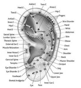

DEFINE AURICULA OF EAR SKIN
Below, the most important and frequent skin diseases of the ear which are potentially subject to surgical or laser therapy are described. In many cases, optimal medical care for patients with skin diseases of the external ear requires an interdisciplinary approach dermatological, ear-nose-throat and surgical collaboration. This is especially true for malignant tumors which may often present at an invasive stage, due to the minimal thickness of the skin compared to other parts of the body. When hidden areas of the outer ear are affected, consultation may be delayed until very late in the disease process.

DEFINE AURICULA OF EAR PROFESSIONAL
Depending on the localization, lesions on the external ear which lead the patient to seek professional help are noticed by the patient himself or by a relative or friend. Further, it has a sound-transmitting function and is located at a visible, esthetically obvious site, drawing considerable attention from the patient. Because of its exposed localization, the ear is particularly liable to the effects of ultraviolet (UV) light and, consequently, to pre-neoplastic and neoplastic skin lesions. The auricle is susceptible to environmental influences and trauma. In case of a ceruminal obstruction, an adequate assessment of the external auditory meatus should be done only after cleaning, which may demask existing dermatosis. The cerumen may mask existing diseases of the skin in the entrance of the external ear canal. An additional anatomical uniqueness is the high concentration of holocrine ceruminal glands in the skin of the external ear canal. In contrast the convex aspect of the outer ear has a thicker subcutis with a stronger layer of subcutaneous fat which causes a certain laxity and displaceability compared to the concave side. The epidermis on the concave aspect lies on a very thin subcutis which is strongly attached to the auricular cartilage. It is attached to the periost and poorly vascularised. Both are of elastic cartilage covered with skin. The outer ear consists of the skin bearing external ear canal and the auricle. When evaluating skin lesions on the ear, specific anatomical peculiarities should be considered.


 0 kommentar(er)
